- They originally defined a cell as a living unit that contained a nucleus. (Now we know about prokaryotic cells, which don't have nuclei -- but it wasn't until much later that this distinction became clear.)
- By the 1850s, it was becoming clear that cells could only be produced by other cells -- omni cellula e cellula, "all cells are from cells", as Rudolf Virchow put it. . .
- Biologists looked at cells in the process of dividing.
- Eukaryote cell division turns out to be a rather complex process. . .
- Normally, a eukaryotic nucleus looks pretty featureless, grainy and uniform.
- There may be a darker-staining spot inside the nucleus -- called the nucleolus -- but that's about it. (More later on what the nucleolus does. . .)
- But when the nucleus is about to divide, the grainy stuff inside appears to "clump", and some small, stringy-looking bodies appear.
- These take up certain stains
very strongly, and so appear strongly colored when the cell is stained.
They're are called by the Greek for "colored bodies," chromosomes.
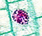
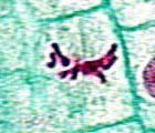
Chromosomes in a cell nucleus at an early stage (left) and a later stage (right) of division
- When cell division begins, each chromosome can be seen to be made up of two long pieces which are stuck together at one point -- looking something like little X's.
- The point where the two pieces join (the center of the "X") -- is called the centromere. . .
- Each of the two pieces that makes up the chromosome is a chromatid (the two halves of the "X"). . .
- Finally, each half of a chromatid is an arm.
- These chromosomes, magnified many thousands of times using a special
microscope, consist of two chromatids and a centromere. Since each chromatid
has two arms, there are four arms in all. (Note that the centromere isn't always
exactly in the center; its position can vary.)
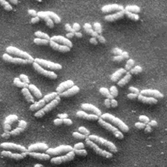
Chromosomes as they look in early cell division, magnified about x10,000.
II. Mitosis
- Mitosis is the name for the complex process of division of the nucleus.
- As mitosis begins, the nuclear membrane disappears. . .
- . . . and the chromosomes move through a series of phases.
- In the first stage of actual division, prophase, the chromosomes
become visible to the light microscope for the first time.
Like so:
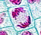
Chromosomes in a cell from an onion root, condensing in preparation for division. - The chromosomes then are arranged in the center of the cell, along an imaginary
plane called the metaphase plate. As you might guess, this stage is called
metaphase. Like so:
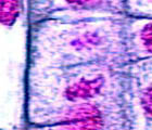
Chromosomes in a cell from an onion root, clustered in the center and about to be pulled apart - Then the chromosomes break apart at the centromere, and each chromatid
is pulled to opposite ends of the dividing nucleus. It's called anaphase.
Like so:
Chromosomes in a cell from an onion root, being pulled apart to opposite ends of the cell - Finally, in what's known as telophase, two new nuclei form. (Usually,
but not always, the cytoplasm is also divided at this time, in a process called
cytokinesis. Either a new cell wall forms between the new nuclei, as in
plants, or the cytoplasm just "pinches" in two, as in animals.) Like so:
Chromosomes in a cell from an onion root, clustering into two new nuclei. The partition line between the two new cells forming is faintly visible.
- In the first stage of actual division, prophase, the chromosomes
become visible to the light microscope for the first time.
Like so:
- Hey, wait a minute! What's dragging these chromosomes around? Answer:
tiny filaments called microtubules.
- During mitosis, clusters of microtubules form at opposite ends of the cell, produced and "directed" by a pair of organelles called centrioles.
- Microtubules and centrioles act as "motors" that do the actual work of pulling chromosomes apart.
- Microtubules are used throughout a cell's life, but when they're specifically
used in mitosis, they are also called spindle fibers.
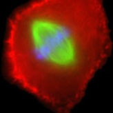
Here the chromosomes have been stained fluorescent blue, and the spindle fibers have been stained green.
By the way, which stage of mitosis is this?
- At the end of cell division, each "daughter cell" has chromosomes consisting of one chromatid each.
- But if you were to follow a cell through interphase and then watch it start to divide again, it would have chromosomes of two chromatids. . .
- Therefore, somewhere in interphase -- somehow which we can't observe directly, at least not with light microscopes -- each chromosome must somehow "grow" a new chromatid which is the same size and shape as the old one.
- Today, we speak of the cell cycle as being divided into four phases:
- The M phase is mitosis -- typical nuclear division, as we described above. . .
- . . . then after division, the cell may grow, move, change shape, etc. etc., making up its first growth phase, or G1 phase. . .
- . . . at some point, the cell's chromosomes develop new chromatids, which is the synthesis phase or S phase. . .
- . . . then the cell may grow, move, change shape some more, etc. etc., making up its second growth phase, or G2 phase. . .
- . . . and then back to mitosis. The G1, S, and G2 phases together are equivalent to interphase.
- Here's a diagram. . .
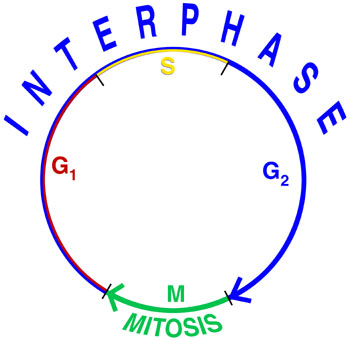
Diagram of the cell cycle