Bone
The vertebrate body is supported by an endoskeleton made of cartilage and bone. (Sharks and their relatives use only cartilage.)
The bones of the human skeleton perform several functions:
- support
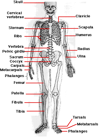
- protection of internal organs from mechanical damage (e.g., skull, ribs)
- reservoir of calcium and phosphate
- source of all the blood cells.
Structure
The diagram below depicts the structure of a typical long bone such as the femur.
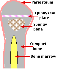
- Periosteum. A tissue covering the bone that brings blood and lymph vessels, as well as nerves, to it
- Compact bone (also known as cortical bone). Dense deposits of minerals — chiefly calcium phosphate — and collagen.
These are arranged in
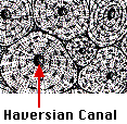 concentric circles around a central Haversian canal through which blood and lymph vessels as well as nerves pass.
- Spongy bone (also known as trabecular or cancellous bone). The mineral deposits are arranged as a system of struts. Bone marrow fill the spaces between.
- Bone marrow. Some bones, such as the femur, also contain a central cavity filled with bone marrow. Bone marrow contains the stem cells that gives rise to all the types of blood cells. [Link]
- Epiphyseal plate. Prior to puberty, this disk of cartilage produces more cartilage which then is converted into more bone. In this way, the bone grows lengthwise. It is stimulated by growth hormone and inhibited by estrogens and progesterone.
Looking at a skeleton, bone seems an inert thing. But in the living body it is anything but.
The size and shape of bones not only changes during growth, but for the rest one's life it is continuously being remodeled in response to the stresses put on it. Approximately 10% of your bone mass is removed and replaced each year.
The remodeling of bone requires the coordinated activity of two types of cells:
- osteoclasts, that demineralize bone in their vicinity
- osteoblasts, that secrete collagen and mineral to lay down new bone in their vicinity.
Osteoclasts are derived from stem cells in the bone marrow — the same ones produce monocytes and macrophages. [Link]
Excess activity of osteoclasts (common after menopause in women) produces osteoporosis. The bones become weakened as cortical bone gets thinner and the spaces in spongy bone get larger.
Reduced activity of osteoclasts produces osteopetrosis. This occurs when a person inherits a mutant version(s) of one or another of the genes needed for normal osteoclast function. Inhibition of osteoclast function causes too much bone to form leading to
- extra-dense bone which actually is more brittle than normal bone thus leading to fractures;
- filling-in of the bone marrow cavity with bone thus interfering with the normal production of blood cells [Link].
Hormones and Bone
In addition to the role of growth hormone (GH) and sex hormones (estrogen and progesterone) in the growth of bones, the remodeling of bone is controlled by:
The links above will take you to descriptions of how these hormones act.
15 September 2005