| Index to this page |
Many tests are now available to detect genetic diseases.
Some examples: Most of these tests can not only be performed on cells removed from adults but also on cells removed fromDuring its development, the fetus sheds cells into the amniotic fluid. After 14–22 weeks of pregnancy, a small volume of this fluid can be removed (using a needle inserted through the abdominal wall).
Separating the cells and culturing them enables the clinician to look forThis is an alternate method of prenatal diagnosis.
A small amount of placental tissue is sucked out by a tube inserted through the abdominal wall or through the vagina (the latter avoiding the need for an incision).
For some tests the fetal cells can be examined immediately without the need to culture them.
Another advantage of CVS is that it can be performed earlier in pregnancy (after only 10–12 weeks) than amniocentesis. If an abortion is to be performed, it is a simpler process early in pregnancy.
This raises the possibility of using genetic tests (e.g., PCR) to identify mutations or chromosomal abnormalities in the fetus. However, only genes contributed by the father (e.g., SRY) can be detected because there is as yet no way to separate the mother's DNA from the fetal DNA. And the tests are not yet as sensitive as amniocentesis and CVS.
Two home blood test kits for determining the sex of the fetus are already on the market. The collected drops of blood are sent to a laboratory to determine whether any Y-chromosome-specific DNA (e.g., SRY) is present.
One of the remarkable facts about mammalian development is that all the cells in the early (e.g., 8-cell) embryo are not needed to produce a healthy fetus (which is why a single fertilized egg can on occasions produce identical twins, triplets, etc.).
So couples using in vitro fertilization (IVF) also can take advantage of genetic screening. While the embryo is in culture, a cell or two can safely be removed and tested for its genotype. For example:
| In mice, at least, a single cell removed from the 8-cell morula can be used to generate a culture of embryonic stem cells. If this turns out to be true for humans, it would provide a method of generating these cells without the need to destroy a blastocyst in the process. |
Thanks to the polymerase chain reaction (PCR), the genotype of an egg can be determined before it is fertilized.
As meiosis I is completed, one set of chromosomes is extruded into the first polar body.
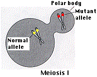
If the mother is heterozygous for a trait, the DNA of the polar body can be amplified by PCR and tested for both alleles.
If the test isFor simplicity, the figure shows only the pair of homologues carrying the locus of concern.
So it is not sufficient to test only for the presence of the mutant gene in the first polar body. You must also demonstrate that the healthy gene is absent. For if crossing over had occurred, the first polar body would contain one mutant and one healthy allele. In that case, a 50:50 chance exists that the other copy of the mutant allele will end up in the egg (instead of in the second polar body).
| Welcome&Next Search |