| Index to this page |
A neuron is a cell specialized to conduct electrochemical impulses called nerve impulses or action potentials.
| Link to a page describing these. |
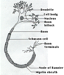
All neurons outside the central nervous system (and many within it) conduct impulses along hairlike cytoplasmic extensions, the nerve fibers or axons. (The diagram represents a motor neuron with most of its axon omitted.) The axons connecting your spinal cord to your foot can be as much as 1 m long (although only a few micrometers in diameter).
Axons grow out of the cell body, which houses the nucleus as well as other organelles such as the endoplasmic reticulum. The length of some axons is so great that it is difficult to see how the cell body can control them. Nevertheless, there is a steady transport of materials (e.g., vesicles, mitochondria) from the cell body along the entire length of the axon. This flow is driven by kinesins moving along the many microtubules in the cytoplasm within the axon.
The function of the axon hillock is described on another page [link to it].
In many neurons, nerve impulses are generated in short branched fibers called dendrites and also in the cell body. The impulses are then conducted along the axon, which usually branches several times close to its end.
Many axons are covered with a glistening fatty sheath, the myelin sheath. It is the greatly-expanded plasma membrane of an accessory cell, the Schwann cell. Schwann cells are spaced at regular intervals along the axon. Their plasma membrane is wrapped around and around the axon forming the myelin sheath.
Where the sheath of one Schwann cell meets that of the next, the axon is unprotected. This region, the node of Ranvier, plays an important part in the propagation of the nerve impulse. [Discussion]
| Link to CNS |
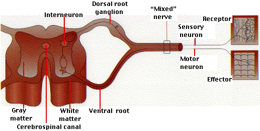
The cell bodies of the sensory neurons leading to the spinal cord are located in clusters, called ganglia, next to the spinal cord. The axons usually terminate at interneurons.
The diagram is a simplified view of the relationship between sensory and motor neurons running to and from the spinal cord.
Interneurons are also called association neurons.
It is estimated that the human brain contains 100 billion (1011) interneurons averaging 1000 synapses on each; that is, some 1014 connections.
The term interneuron hides a great diversity of structural and functional types of cells. In fact, it is not yet possible to say how many different kinds of interneurons are present in the human brain. Certainly hundreds; perhaps many more.
Most motor neurons are stimulated by interneurons, although some are stimulated directly by sensory neurons.
| View an example |
| Welcome&Next Search |