Cells contain elaborate arrays of protein fibers that serve such functions as:
- establishing cell shape
- providing mechanical strength
- locomotion
- chromosome separation in mitosis and meiosis
- intracellular transport of organelles
The cytoskeleton is made up of three kinds of protein filaments:
- Actin filaments (also called microfilaments)
- Intermediate filaments and
- Microtubules
Monomers of the protein actin polymerize to form long, thin fibers. These are about 8 nm in diameter and, being the thinnest of the cytoskeletal filaments, are also called microfilaments. (In skeletal muscle fibers they are called "thin" filaments.)
Some functions of actin filaments:
- form a band just beneath the plasma membrane that
- provides mechanical strength to the cell
- links transmembrane proteins (e.g., cell surface receptors) to cytoplasmic proteins
- anchors the centrosomes at opposite poles of the cell during mitosis
- pinches dividing animal cells apart during cytokinesis
- generate cytoplasmic streaming in some cells
- generate locomotion in cells such as white blood cells and the amoeba
- interact with myosin ("thick") filaments in skeletal muscle fibers to provide the force of muscular contraction
These cytoplasmic fibers average 10 nm in diameter (and thus are "intermediate" in size between actin filaments (8 nm) and microtubules (25 nm)(as well as of the thick filaments of skeletal muscle fibers).
There are several types of intermediate filament, each constructed from one or more proteins characteristic of it.
- keratins are found in epithelial cells and also form hair and nails;
- nuclear lamins form a meshwork that stabilizes the inner membrane of the nuclear envelope;
- neurofilaments strengthen the long axons of neurons;
- vimentins provide mechanical strength to muscle (and other) cells.
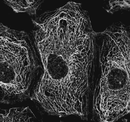
Despite their chemical diversity, intermediate filaments play similar roles in the cell: providing a supporting framework within the cell. For example, the nucleus in epithelial cells is held within the cell by a basketlike network of intermediate filaments made of keratins. (photo at right)
In the photo (courtesy of W. W. Franke), a fluorescent stain has been used to show the intermediate filaments of keratin in epithelial cells.
Different kinds of epithelia use different keratins to build their intermediate filaments.
Over 20 different kinds of keratins have been found, although each kind of epithelial cell may use no more than 2 of them. Up to 85% of the dry weight of squamous epithelial cells can consist of keratins.
 Microtubules
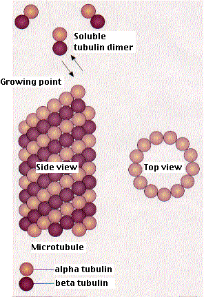
- are straight, hollow cylinders
- have a diameter of about 25 nm
- are variable in length but can grow 1000 times as long as they are thick
- are built by the assembly of dimers of alpha tubulin and beta tubulin.
- are found in both animal and plant cells
Microtubules - grow at each end by the polymerization of tubulin dimers (powered by the hydrolysis of GTP), and
- shrink at each end by the release of tubulin dimers (depolymerization)
However, both processes always occur more rapidly at one end, called the plus end. The other, less active, end is the minus end.
Microtubules participate in a wide variety of cell activities. Most involve motion.
The motion is provided by protein "motors" that use the energy of ATP to move along the microtubule.
There are two major groups of microtubule motors:
- kinesins (most of these move toward the plus end of the microtubules) and
- dyneins (which move toward the minus end).
Some examples:
- The rapid transport of organelles, like vesicles and mitochondria, along the axons of neurons takes place along microtubules with their plus ends pointed toward the end of the axon. The motors are kinesins.
| Charcot-Marie-Tooth disease. One cause of this rare disorder is an inherited mutated gene for one of the kinesins. In these patients, axonal transport is defective (which probably accounts for their muscle weakness first occurring in muscles at the ends of the longer motor neurons). |
- The migration of chromosomes in mitosis and meiosis takes place on microtubules that make up the spindle fibers. Both kinesins and dyneins are used as motors as we shall see below.
In plant cells, microtubules are created at many sites scattered through the cell. In animal cells, the microtubules originate at the centrosome.
 The centrosome is
- located in the cytoplasm attached to the outside of the nucleus.
- Just before mitosis, the centrosome duplicates.
- The two centrosomes move apart until they are on opposite sides of the nucleus.
- As mitosis proceeds, microtubules grow out from each centrosome with their plus ends growing toward the metaphase plate. These clusters of microtubules are called spindle fibers.
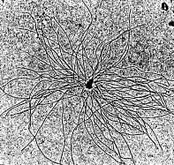
The photo (courtesy of Tim Mitchison) shows microtubules growing in vitro from an isolated centrosome. The centrosome was supplied with a mixture of alpha and beta tubulin monomers. These spontaneously assembled into microtubules only in the presence of centrosomes.
Spindle fibers have three destinations:
- Some attach to one kinetochore of a dyad with those growing from the opposite centrosome binding to the other kinetochore of that dyad.
- Some bind to the arms of the chromosomes.
- Still others continue growing from the two centrosomes until they extend between each other in a region of overlap.
All three groups of spindle fibers participate in
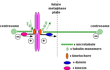
- the assembly of the chromosomes at the metaphase plate at metaphase. Proposed mechanism (the diagram shows only 1 and 2):
- Microtubules attached to opposite sides of the dyad shrink or grow until they are of equal length.
- Microtubules motors attached to the kinetochores move them
- toward the minus end of shrinking microtubules (a dynein);
- toward the plus end of lengthening microtubules (a kinesin).
- The chromosome arms use a different kinesin to move to the metaphase plate.
- the separation of the chromosomes at anaphase.
- The sister kinetochores separate and, carrying their attached chromatid,
- move along the microtubules powered by minus-end motors, dyneins, while the microtubules themselves shorten (probably at both ends).
- The overlapping spindle fibers move past each other (pushing the poles farther apart) powered by plus-end motors, the "bipolar" kinesins.
- In this way the sister chromatids end up at opposite poles.
Other Functions of Centrosomes
In addition to their role in spindle formation, centrosomes play other important roles in animal cells:
- signaling that it is o.k. to proceed to cytokinesis. Destruction of both centrosomes with a laser beam prevents cytokinesis even if mitosis has been completed normally.
- signaling that it is o.k. for the daughter cells to begin another round of the cell cycle; specifically to duplicate their chromosomes in the next S phase. Destruction of one centrosome with a laser beam still permits cytokinesis but the daughter cells fail to enter a new S phase.
- Segregating signaling molecules (e.g., mRNAs) so that they pass into only one of the two daughter cells produced by mitosis. In this way, the two daughter cells can enter different pathways of differentiation even though they contain identical genomes. [Link to further discussion.]
- In at least some developing neurons, the position of the centrosome establishes the point at which the axon will grow out.
Cancer cells often have more than the normal number (1 or 2 depending on the stage of the cell cycle) of centrosomes . They also are aneuploid (have abnormal numbers of chromosomes), and considering the role of centrosomes in chromosome movement, it is tempting to think that the two phenomena are related.
Mutations in the tumor suppressor gene p53 seem to predispose the cell to excess replication of the centrosomes.
Chromosome movement in mitosis also involves polymerization and depolymerization of the microtubules. Taxol, a drug found in the bark of the Pacific yew, prevents depolymerization of the microtubules of the spindle fiber. This, in turn, stops chromosome movement, and thus prevents the completion of mitosis. Taxol is being used with some success as an anticancer drug.
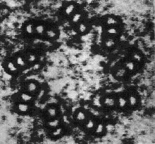 Each centrosome contains a pair of centrioles.
Centrioles are built from a cylindrical array of 9 microtubules, each of which has attached to it 2 partial microtubules.
The photo (courtesy of E. deHarven) is an electron micrograph showing a cross section of a centriole with its array of nine triplets of microtubules. The magnification is approximately 305,000.
When a cell enters the cell cycle, and proceeds from G1 to S phase, each centriole is duplicated. A "daughter" centriole grows out of the side of each parent centriole. Thus centriole replication — like DNA replication (which is occurring at the same time) — is semiconservative.
Once formed, most of the functions of the centrosomes can be accomplished without centrioles. However,
- Centrioles appear to be needed to organize the centrosome in which they are embedded.
- Sperm cells contain a pair of centrioles; eggs have none. The sperm's centrioles are absolutely essential for forming a centrosome which will form a spindle enabling the first division of the zygote to take place.
- Centrioles are also needed to make cilia and flagella.
Cilia and Flagella
Both cilia and flagella are constructed from microtubules, and both provide either
- locomotion for the cells (e.g., sperm) or
- move fluid past the cells (e.g., ciliated epithelial cells that line our air passages and move a film of mucus towards the throat).
Both cilia and flagella have the same basic structure. If the cell has
- many short ones, we call them cilia or
- only one or a few long ones, we call them flagella.
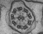 Each cilium (or flagellum) is made of
- a cylindrical array of 9 evenly-spaced microtubules, each with a partial microtubule attached to it. This gives the structure a "figure 8" appearance when view in cross section.
- 2 single microtubules run up through the center of the bundle, completing the so-called "9+2" pattern.
- The entire assembly is sheathed in a membrane that is simply an extension of the plasma membrane.
This electron micrograph (courtesy of Peter Satir) shows the 9+2 pattern of microtubules in a single cilium seen in cross section.
Motion of cilia and flagella is created by the microtubules sliding past one another — Link. This requires:
- motor molecules of dynein, which link adjacent microtubules together, and
- the energy of ATP.
Each cilium or flagellum grows out from, and remains attached to, a basal body embedded in the cytoplasm. Basal bodies are identical to centrioles and are, in fact, produced by them.
13 August 2005

 Microtubules
 The centrosome is

 Each centrosome contains a pair of centrioles.
 Each cilium (or flagellum) is made of