Cilia and Flagella
These whiplike appendages extend from the surface of many types of eukaryotic cells.
- If there are many of them, they are called cilia;
- if only one, or a few, they are flagella. Flagella also tend to be longer than cilia but are otherwise similar in construction.
Function
Cilia and flagella move liquid past the surface of the cell.
- For single cells, such as sperm, this enables them to swim.
- For cells anchored in a tissue, like the epithelial cells lining our air passages, this moves liquid over the surface of the cell (e.g., driving particle-laden mucus toward the throat).
Structure
Both cilia and flagella consist of:
- a cylindrical array of 9 filaments consisting of:
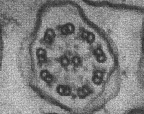
- a complete microtubule extending into the tip of the cilium;
- a partial microtubule that doesn't extend as far into the tip.
- cross-bridges of the motor protein dynein that extend from the complete microtubule of one filament to the partial microtubule of the adjacent filament.
- a pair of single microtubules running up through the center of the bundle, producing the "9+2" arrangement.
- The entire assembly is sheathed in a membrane that is an extension of the plasma membrane.
This electron micrograph (courtesy of Peter Satir) shows a cilium in cross section.
Each cilium (and flagellum) grows out from, and remains attached to, a basal body embedded in the cytoplasm. Basal bodies are identical to centrioles and are, in fact, produced by them. For example, one of the centrioles in developing sperm cells — after it has completed its role in the distribution of chromosomes during meiosis — becomes a basal body and produces the flagellum.
The bending of cilia (and flagella) has many parallels to the contraction of skeletal muscle fibers.
In the case of cilia and flagella, dynein powers the sliding of the microtubules against one another — first on one side, then on the other.
Testing the Model
- Remember: the partial microtubules do not extend as far into the tip as the complete microtubules.
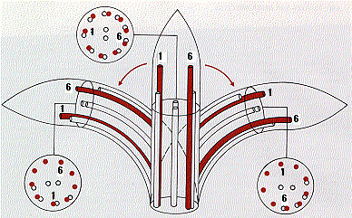
- So if a slice is made a short distance back from the tip,
- A straight cilium should show the complete pattern (center of diagram).
- In a bent cilium, approximately half the filaments on the upper side should be retracted because of the greater arc on the convex side. So the partial microtubules would disappear being drawn below the plane of the slice. As seen here, bending to the left causes the partial microtubules 4, 5, 6, 7, and 8 to disappear.
- When the cilium bends the other way, the partial microtubules on the opposite side disappear while they reappear on what is now the lower or concave side.
- Electron micrographs (made by Peter Satir) have verified this model precisely.
Other Parallels
There are other parallels between the sliding filaments of skeletal muscle and the sliding microtubules of cilia.
- Both are powered by ATP.
- Dynein (like myosin) is the ATPase.
- Both are regulated by calcium ions.
Motile, "9+2", cilia are found only on certain cells in the vertebrate body, e.g., the epithelia lining the airways.
But almost every cell in vertebrates has — or had — a single primary cilium.
These do not beat because they lack the central pair of microtubules; that is they are "9+0".
Where functions have been identified, they all involve sensory reception.
Some examples:
Mechanoreceptors
A primary cilium extends from the apical surface of the epithelial cells lining the kidney tubules and monitors the flow of fluid through the tubules. Inherited defects in the formation of these cilia cause polycystic kidney disease.
Chemoreceptors
We detect odors by receptors on the primary cilium of olfactory neurons. [Link]
Photoreceptors
The outer segment of the rods in the vertebrate retina is also derived from a primary cilium. [View]
22 October 2005