These diseases
- are transmissible — from host to host of a single species and, sometimes, even from one species to another (such as a laboratory animal)
- destroy brain tissue giving it a spongy appearance
For these reasons, prion diseases are also called transmissible spongiform encephalopathies or TSEs.
Some examples:
Before the victim dies of a TSE, the damage to the brain is reflected in such signs as loss of coordination and — in humans — dementia.
Injections of ground-up brain tissue from an animal or human patient with a prion disease into another animal (of the appropriate species) transmits the disease. This suggests that the disease is caused by an infectious agent such as a virus. But viruses have a genome and — despite intense efforts — no evidence of a virus has ever been found in these brain extracts. In fact, treating the extracts with agents (e.g., ultraviolet light) that destroy DNA does not reduce their infectiousness.
To date, the evidence indicates that the infectious agent in the TSEs is a protein.
Stanley Prusiner who
- pioneered in the study of these proteins and
- was awarded the Nobel Prize in 1997 for his efforts
has named them prion proteins (designated PrP) or simply prions.
It turns out that prions are molecules of a normal body protein that have changed their three-dimensional configuration.
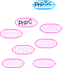
The normal protein
- is called PrPC (for cellular)
- is a glycoprotein normally found at the cell surface inserted in the plasma membrane
- has its secondary structure dominated by alpha helices (probably 3 of them)
- is easily soluble
- is easily digested by proteases
- is encoded by a gene designated (in humans) PRNP located on our chromosome 20.
The abnormal, disease-producing protein
- is called PrPSc (for scrapie)
- has the same amino acid sequence as the normal protein; that is, their primary structures are identical but
- its secondary structure is dominated by beta conformation
- is insoluble in all but the strongest solvents
- is highly resistant to digestion by proteases
- When PrPSc comes in contact with PrPC, it converts the PrPC into more of itself (even in the test tube).
- These molecules bind to each other forming aggregates.
- It is not yet clear if these aggregates are themselves the cause of the cell damage or are simply a side effect of the underlying disease process.
10–15% of the cases of CJD are inherited; that is, the patient comes from a family in which the disease has appeared before.
The disease is inherited as an autosomal dominant.
The patients have inherited at least one copy of a mutated PRNP gene. Some of the most common mutations are:
- a change in codon 200 converting glutamic acid (E) at that position to lysine (K)(thus designated "E200K")[link to a table giving the single-letter code for the amino acids]
- a change from aspartic acid (D) at position 178 in the protein to asparagine (D178N) when it is accompanied by a polymorphism in the gene encoding valine at position 129 . (When the polymorphism at codon 129 is Met/Met, the D178N mutation produces Fatal Familial Insomnia instead.)
- a change from valine (V) at position at position 210 to isoleucine (V210I)
Extracts of autopsied brain tissue from these patients can transmit the disease to
- apes (whose PRNP gene is probably almost identical to that of humans).
- transgenic mice who have been given a Prnp gene that contains part of the human sequence.
These results lead to the important realization that prion diseases can only be transmitted to animals that already carry a PRNP gene with a sequence that is at least similar to the one that encoded the PrPSc. In fact, knockout mice with no Prnp genes at all cannot be infected by PrPSc.
This prion disease is caused by the inheritance of a PRNP gene with a mutations encoding most commonly
- leucine instead of proline at position 102 (P102L) or
- valine instead of alanine at position 117 (A117V)
Again, the disease is also strongly associated with homozygosity for a polymorphism at position 129 (both residues being methionine).
Brain extracts from patients with GSS can transmit the disease to
- monkeys and apes
- transgenic mice containing a portion of the human PRNP gene.
Transgenic mice expressing the P102L gene develop the disease spontaneously.
People with this rare disorder have inherited
- a PRNP gene with asparagine instead of aspartic acid encoded at position 178 (D178N)
- the susceptibility polymorphism of methionine at position 129 of the PRNP gene.
Extracts from autopsied brains of FFI victims can transmit the disease to transgenic mice.
Kuru was once found among the Fore tribe in Papua New Guinea whose rituals included eating the brain tissue of their recently deceased members of the tribe. Since this practice was halted, the disease has disappeared.
Before then, the disease was studied by transmitting it to chimpanzees using injections of autopsied brain tissue from human victims.
This disease of sheep (and goats) was the first TSE to be studied. It seems to be transmitted from animal to animal in feed contaminated with nerve tissue. It can also be transmitted by injection of brain tissue.
An epidemic of this disease began in Great Britain in 1985 and before it was controlled, over 170,000 cattle were sickened by it. Its origin appears to have been cattle feed that
- contained brain tissue from sheep infected with scrapie and
- had been treated in a new way that no longer destroyed the infectiousness of the scrapie prions.
The use of such food was banned in 1988 and after peaking in 1992, the epidemic declined quickly.
A number of humans have acquired CJD through accidental exposure to material contaminated with CJD prions.
- Grafts of dura mater taken from patients with inherited CJD have transmitted the disease to recipients.
- Corneal transplants have also inadvertently transmitted CJD.
- Instruments used in brain surgery on patients with CJD have transmitted the disease to other patients. Two years after their supposed sterilization, these instruments remained infectious.
- Over 100 people have acquired CJD from injections of human growth hormone or human gonadotropins prepared from pooled pituitary glands that inadvertently included glands taken from humans with CJD.
Now that both hGH and human gonadotropins are available through recombinant DNA technology, such disastrous accidents need never recur.
This human disorder appeared some years after the epidemic of BSE (Mad Cow Disease) swept through the cattle herds in Great Britain. Even though the cow and human PRNP genes differ at 30 codons, the sequence of their prions suggests that these patients (155 by 2005) acquired the disease from eating contaminated beef.
All the patients are homozygous for the susceptibility polymorphism of methionine at position 129.
The BSE epidemic has waned, and slaughter techniques that allow cattle nervous tissue in beef for human consumption have been banned since 1989. Now we must wait to see whether more cases of vCJD are going to emerge or whether the danger is over.
A number of TSEs have been found in other animals.
- Cats are susceptible to Feline Spongiform Encephalopathy (FSE)
- Mink are also susceptible to a TSE.
- Even though mad cow disease has not been seen in North America, a similar disease is found in elk and mule deer in the Rocky Mountains of the U.S.
CJD and FFI occasionally occur in people who have no family history of the disease and no known exposure to infectious prions.
The cause of their disease is uncertain.
- Perhaps a spontaneous somatic mutation has occurred in one of the PRNP genes in a cell.
- Perhaps their normal PrPC protein has spontaneously converted into the PrPSc form.
Whatever the answer, all the cases are found in people with a susceptibility polymorphism in their PRNP genes.
Two proteins in yeast (Saccharomyces cerevisiae)
- the Sup35 protein ("Sup35p") and
- the Ure2 protein (Ure2p)
are able to form prions; that is, they can exist either
- in a PrPC-like form that is functional or
- in a PrPSc-like form that is not.
The greater ease with which yeast can be studied has
- proved that only protein is involved in prion formation and
- provided insight into the need for PrPSc to find PrPC molecules of a similar primary structure in order to be able to convert them into the PrPSc form.
- A particular PrPSc can only convert PrPC molecules of the same — or at least similar — primary structure.
- This requirement of "like-with-like" resides in a short sequence at the N-terminal of the protein (rather like an antibody epitope).
- Yeasts engineered to form two types of prion form two types of "pure" aggregates within the cell.
- Even in the test tube, each type of prion finds and aggregates with others of its own type.
 So the picture that emerges is that a molecule of PrPSc
- acts as a "seed" providing a template for converting PrPC to more PrPSc
- These interact with each other to form aggregates.
Although only a small portion of the prion protein is responsible for its specificity, other parts of the molecule are needed for flipping the molecule from the alpha-helical to the beta conformation. All prion proteins contain tracts of repeated Gln-Asn residues which appear to be essential for the conversion process.
The deposits of PrPScin the brain are called amyloid. Amyloid deposits are also found in other diseases involving the brain, such as Alzheimer's disease.
Most cells, including neurons in the brain, contain proteasomes that are responsible for degrading misfolded or aggregated proteins. In the various brain diseases characterized by a build-up of amyloid deposits, it appears that the amount of amyloid overwhelms the capacity of the proteasomes to do their job. Because of the critical role of proteasomes in other cell functions, such as mitosis, it is easy to see why these deposits lead to death of the cell.
Evidence:
- Yeast are not harmed when Sup35p and Ure2p form prions.
- The role of CPEB.
CPEB ("cytoplasmic polyadenylation element binding protein") is a protein that
Recent evidence (from the same lab that studies LTF in Aplysia — Link) indicates that this CPEB
- accumulates at activated (by serotonin) synapses;
- has the ability to undergo a change in tertiary structure that
- persists for long periods;
- induces the same conformational change in other molecules of CPEB forming prion-like aggregates.
Perhaps the accumulation of these aggregates at a stimulated synapse causes a long-term change in its activity (memory).
5 May 2005