Helper T cells are
There are two distinct kinds:
- Th1
- Th2
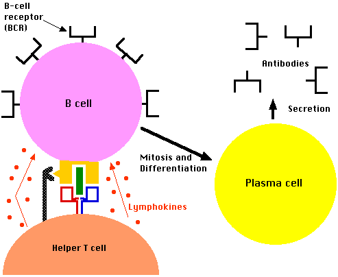
- These provide help for B cells and, in so doing, are essential for antibody-mediated immunity. Antibodies are needed to control extracellular pathogens (which — unlike intracellular parasites — are exposed to antibodies in blood and other body fluids).
Like all T cells, Th cells arise in the thymus.
- When they acquire CD4, they are called pre-Th cells.
- When they are presented with both
they begin to proliferate and become activated.
- It is the nature of the stimulation that determines which path they enter: the path leading to Th1 cells or the path leading to Th2 cells.
The antigen-presenting cells (APCs) are called dendritic cells (DC). They
(Dendritic cells can also present intact antigen directly to B cells. In this case, the engulfed antigen is not degraded in lysosomes but is returned to the cell surface for presentation to B cells bearing BCRs of the appropriate specificity.)
There are two kinds of dendritic cell that present antigens to T cells:
- DC1 — these are descended from monocytes
- DC2 — these appear to be derived from lymphocytes
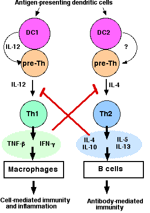 Th1 cells are produced when DC1-type dendritic cells and pre-Th cells form an immunological synapse in which the dendritic cell
- presents antigen to the T cell's receptor for antigen (TCR);
- secretes interleukin 12 (IL-12).
The paracrine stimulation by IL-12 activates (through JAK-STAT pathways) the Th1 cells to secrete their own lymphokines:
- tumor-necrosis factor-beta (TNF-β) (also known as lymphotoxin) and
- interferon-gamma (IFN-γ)
These
- stimulate macrophages to kill the bacteria they have engulfed;
- recruit other leukocytes to the site producing inflammation.
Th2 cells are produced when DC2-type dendritic cells present antigen to the T cell's receptor for antigen (TCR) and, presumably, one or more paracrine stimulants. The identity of the cytokine(s) is still uncertain (indicated by a ? in the figure).
The major lymphokines secreted by Th2 cells are
- interleukin 4 (IL-4). This
- stimulates class-switching in B cells and promotes their synthesis of IgE antibodies.
- acts as a positive-feedback device promoting more pre-Th cells to enter the Th2 pathway.
- blocks the IFN-γ receptors from entering the immunological synapse on pre-Th cells thus inhibiting them from entering the Th1 path (shown in red).
- Interleukin 5 (IL-5). Attracts and activates eosinophils.
- Interleukin 10 (IL-10). Inhibits IL-12 production by DCs thus inhibiting pre-Th cells from entering the Th1 pathway.
- Interleukin 13 (IL-13). This also promotes the synthesis of IgE antibodies.
Two transcription factors have been found that play a critical role in the choice between becoming a Th1 or a Th2 cell.
- T-bet for Th1 cells
- GATA-3 for Th2 cells
T-bet produces Th1 cells by
- turning on the genes needed for Th1 function (e.g., for IFN-γ)
- blocking the activity of GATA-3.
Mice whose genes for T-bet have been "knocked-out" lack Th1 cells and have elevated numbers of Th2 cells (making them susceptible to such Th2-mediated disorders as asthma).
|
The CD8+ Cytotoxic T Cells (CTL) also come in two subsets:
- Tc1 that, like Th1 cells, secrete IFN-γ and
- Tc2 that, like Th2 cells, secrete IL-4.
|
The antigenic stimulus that sends pre-Th cells down one path or the other also sets the stage for reinforcing the response.
A Th1 response inhibits the Th2 path in two ways:
- IFN-γ
(shown above in red) and IL-12 inhibit the formation of Th2 cells;
- IFN-γ
also inhibits class-switching in B cells.
A Th2 response inhibits the Th1 path:
- IL-4 and IL-10 suppress Th1 formation (shown above in red).
There is also evidence that late in the immune response, negative feedback mechanisms come into play to dampen the response.
- IL-4 kills the precursors of the DC2 cells (by apoptosis) thus inhibiting the Th2 path and further production of IL-4
- IFN-γ
may eventually turn off the Th1 response that produced it.
Chemokines are cytokines that are chemotactic for (attract) leukocytes. Because they are chemotactic cytokines, chemokines are designated by the initials CC.
Chemokines bind to receptors on the responding leukocyte. The receptors are transmembrane proteins with the chemokine binding site exposed at the surface of the plasma membrane. Chemokine receptors are designated CCR.
With their different functions, we might expect that Th1 cells and Th2 cells would respond differently to chemokines. And so they do.
- Th1 cells express the chemokine receptor CCR5 (but not CCR3).
- Th2 cells express the chemokine receptor CCR3 (but not CCR5).
CCR3
One chemokine that binds to CCR3 is called eotaxin.
It is secreted by epithelial cells and phagocytic cells in regions where allergic reactions are occurring.
CCR3 is found on
all cells implicated in allergic responses (e.g., asthma).
CCR5 is found on
- Th1 cells, especially those in the lymphoid tissue of the intestine
- macrophages
CCR5 also acts — along with the CD4 molecule — as a coreceptor for HIV-1, the retrovirus that causes AIDS. This fact may explain
- why destruction of the lymphoid tissue of the intestine occurs soon after HIV infection;
- why certain HIV-infected men
- with inherited mutations in CCR5 or
- who produce high levels of the natural ligand for CCR5 (a chemokine designated CCL3L1). CCL3L1 presumably competes with HIV for access to CCR5.
can tolerate their infection for long periods without progressing to a full-blown case of AIDS;
- the collapse of cell-mediated immunity in the late stages of AIDS.
4 December 2005